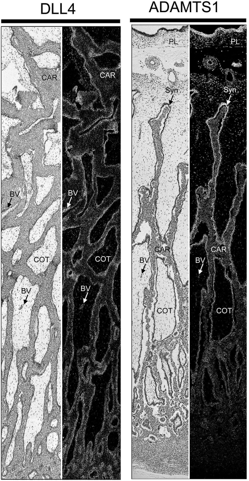
FIG. 10.
Composite images of in situ hybridization analyses for DLL4 and ADAMTS1 mRNA within placentomes collected from unilaterally pregnant ewes on Day 80 of pregnancy. Corresponding bright field and dark field images of representative placentomes are shown. BV, blood vessel; CAR, caruncular tissue; COT, cotyledonary tissue; PL, chorion; Syn, epithelial syncytia of the placentome. Representative images are shown from independent samples (n = 4). Width of each field is 940 μm.

