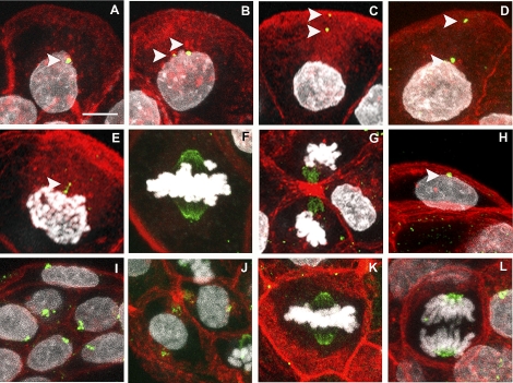
FIG. 1.
Localization of TUBG1 in rat pre-implantation embryos. Eight- to 16-cell embryos (A–G) and blastocysts (H–J) were stained for visualization of TUBG1 (green), f-actin (red), and DNA (white). At the 8- to 16-cell stage, interphase blastomeres exhibited one or two TUBG1 foci with perinuclear or cortical positioning (A–D, arrows). TUBG1 was also localized to spindle poles and midbodies of mitotic blastomeres (E–G). In blastocysts, aggregates of TUBG1 were observed in TE (H) and ICM (J) cells and at the spindle at metaphase (K) and chromosomes at anaphase (L). Bar = 10 μm.

