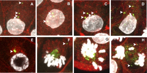
FIG. 2.
Localization of TUBG1 in rat preimplantation embryos following chronic maternal TCDD exposure. Eight- to 16-cell embryos (A–H) were collected from female rats chronically exposed to TCDD and stained for visualization of TUBG1 (green), f-actin (red), and DNA (white). Interphase blastomeres exhibited multiple TUBG1 foci (A–D, arrows). Some prophase blastomeres only had one foci (E). TUBG1 was also localized to a single spindle pole at metaphase, with foci detected in the cytoplasm but not contributing to the spindle (F, arrow). In some mitotic blastomeres, appropriate chromosome alignment (G) and segregation failed (H). Bar = 10 μm.

