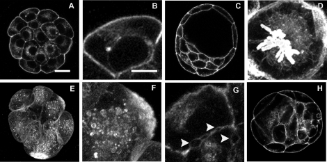
FIG. 5.
Maternal TCDD exposure modifies f-actin organization in preimplantation embryos. Eight- to 16-cell embryos (A, B, and D–G) and blastocysts (C and H) were collected from control female rats (A–D) and rats exposed chronically (E–G) or acutely (H) to TCDD and stained for visualization f-actin. TCDD treatment resulted in cytoplasmic vesicles and f-actin aggregates (E and F), vacuoles (H), and disruption of adherence properties between blastomeres (G, arrows). These features were not apparent in control embryos (A–H). Bar = 20 μm (A, C, E, and H) or 10 μm (B, D, F, and G).

