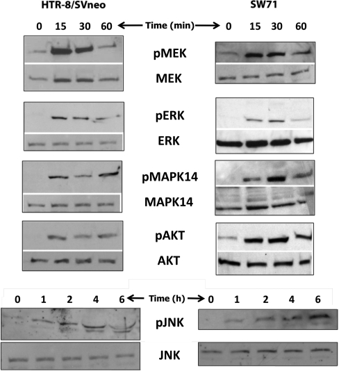
FIG. 1.
Identification of signaling pathways activated by HBEGF. Extracts were prepared from HTR-8/SVneo (left panels) or SW.71 (right panels) cell lines at the indicated times after treatment with 1 nM HBEGF and analyzed by Western blotting. Each lane contained 30 μg of protein extract and was labeled with antibodies against the indicated proteins (lower panels) or their phosphorylated forms (upper panels). Images shown are representative of at least three experiments.

