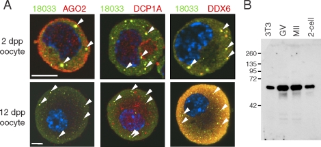
FIG. 1.
P-bodies are present in small incompetent oocytes. A) Confocal images of mouse meiotically incompetent oocytes from 2 dpp and 12 dpp females after staining with 18033, AGO2, DCP1A, and DDX6 antibodies. Staining with 18033 is green colored, other proteins are shown in red, and DNA staining (DAPI) is shown in blue. Colocalization yields yellow color. The arrowheads depict P-bodies. For each sample, at least 10 oocytes were examined, and representative images are shown. Bar = 10 μm. B) DDX6 antibody specificity analyzed by Western blot analysis. DDX6 antibody used in this work detects a single band corresponding to the size of DDX6 (RCK/p54).

