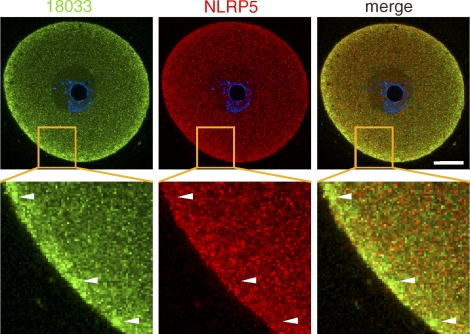
FIG. 5.
Confocal images showing immunolocalization of 18033 and NLRP5 in GV SN oocytes. Identities of green and red channels are indicated above each panel, and colocalization yields yellow color. The blue signal is DNA staining (DAPI). The area in the merged image outlined by solid lines is magnified in the adjacent image. The arrowheads point to subcortical RNP aggregates. Ten oocytes were examined, and representative images are shown. SN, surrounded nucleolus GV oocyte; NSN, nonsurrounded nucleolus GV oocyte. Bar = 20 μm.

