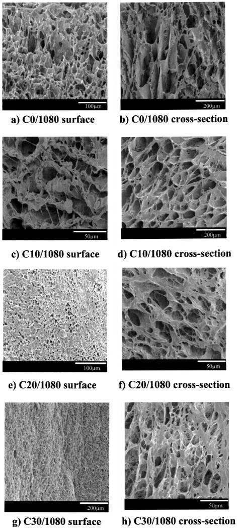
Figure 1.

Scanning electron micrographs of PEUU/collagen scaffolds generated by varying the PEUU/collagen ratio. (a, b) 100:0 PEUU/collagen, (c, d) 90:10; (e, f) 80:20; (g, h) 70: 30. For polymer notation refer to Table 1. Scaffold surface morphologies are seen in (a), (c), (e), and (g). Cross-sectional morphologies are seen in (b), (d), (f), and (h). All scaffolds were prepared using a 10% PEUU solution and a quenching temperature of −80°C. Scale bars are presented in the lower right corner for each image.
