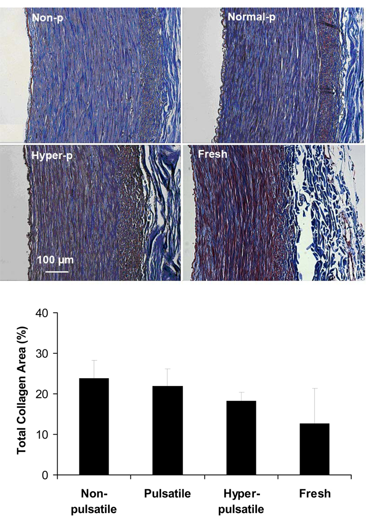
Figure 3.
Top: Light microscope images of trichrome stained porcine carotid arteries cultured under nonpulsatile pressure (non-p), normal pulsatile pressure (normal-p), and hyperpulsatile pressure (hyper-p) for 7 days as compared to a fresh artery. The trichrome stain colors collagen blue and elastin black. Bottom: Percent area of collagen calculated using photometric measurement in arteries cultured under different pulse pressures. Data were plotted as means± SD. Sample size n=3, 4, 6, and 5, respectively.

