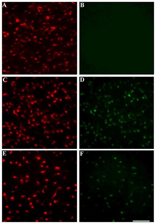
Figure 1. gp120 injected into the superior colliculus is retrogradely transported to RGC.

Rats (n=4 each group) were microinjected with FR and heat-inactivated gp120 (control) or FR and gp120 and sacrificed 3 or 18 days later. RGC were analyzed for FR (A, C and E) and gp120 immunoreactivity (B, D and F). A and B, control rats; C and D, rats sacrificed 3 days after the injection of gp120; E and F, rats sacrificed 18 days after gp120 injection. Images are representative of seventy two microscopic fields of retina per animal. By 3 days post-treatment, 35 ± 6 cells per field were gp120 positive (equivalent to ~76% of FR positive cells). By 18 days, 22 ± 7 cells per field were gp120 positive (~48% of FR positive cells). Scale bar = 100μm.
