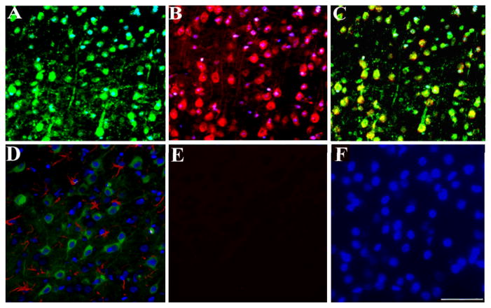
Figure 3. Transport of gp120 occurs in neurons but not in astrocytes.
Rats were microinjected with gp120 into the superior colliculus. Three days later, rats were sacrificed and gp120 immunoreactivity was analyzed in serial sections from the visual cortex. A, example of a section stained for neurofilament (green) and DAPI (blue). B, example of a section stained for gp120 (red) and DAPI (blue). C, merge of A and B. D, example of a section stained for GFAP (red), DAPI (blue) and gp120 (green). E, example of a section in which the primary antibody for gp120 was omitted. F, section in E was counterstained for DAPI (blue). Images are representative of ten sections per animal. Scale bar= 100μm.

