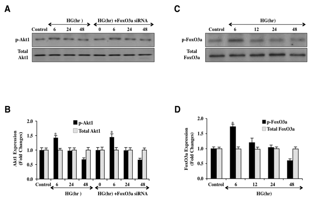
Fig. 2. Elevated D-glucose results in the eventual loss of phosphorylation of Akt1 and the increased activity of FoxO3a.
In A and B, primary EC protein extracts (50 µ/lane) were immunoblotted with anti-phosphorylated-Akt1 (p-Akt1, Ser473) or anti-total Akt1 at 0 (control), 6, 24, and 48 hours following administration of elevated D-glucose (HG = high glucose, 20 mM). Phosphorylated (active) Akt1 (p-Akt1) expression is initially increased at 6 hours following elevated D-glucose, but subsequently is reduced and returns to control levels at 24 hours and 48 hours after elevated D-glucose (*P<0.01 vs. control). Total Akt1 is not altered. In C and D, primary EC protein extracts (50 µ/lane) were immunoblotted with anti-phosphorylated-FoxO3a (p-FoxO3a, Ser253) or anti-total FoxO3a at 0, 6, 24, and 48 hours following administration of elevated D-glucose (HG = high glucose, 20 mM). Correlating with Akt1 activity, active (unphosphorylated) FoxO3a (p-FoxO3a) expression is increased at 12, 24, and 48 hours after elevated D-glucose but total FoxO3a is not affected (*P<0.01 vs. control).

