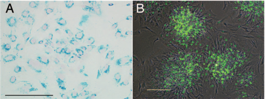
Figure 1.
In vitro characterization of labeled rat MSCs. A, Prussian blue staining of magnetically labeled cells. Nearly 100% of the cells are efficiently labeled with a characteristic perinuclear, endosomal distribution of the Feridex iron (blue precipitate). B, Fluorescent photomicrograph of anti-BrdU immunostaining, merged with phase-contrast image. BrdU-positive nuclei (green) are indicative of proliferative cells in the form of colonies, whereas peripheral cells are nondividing. Scale bars in A and B equal 200 µm.

