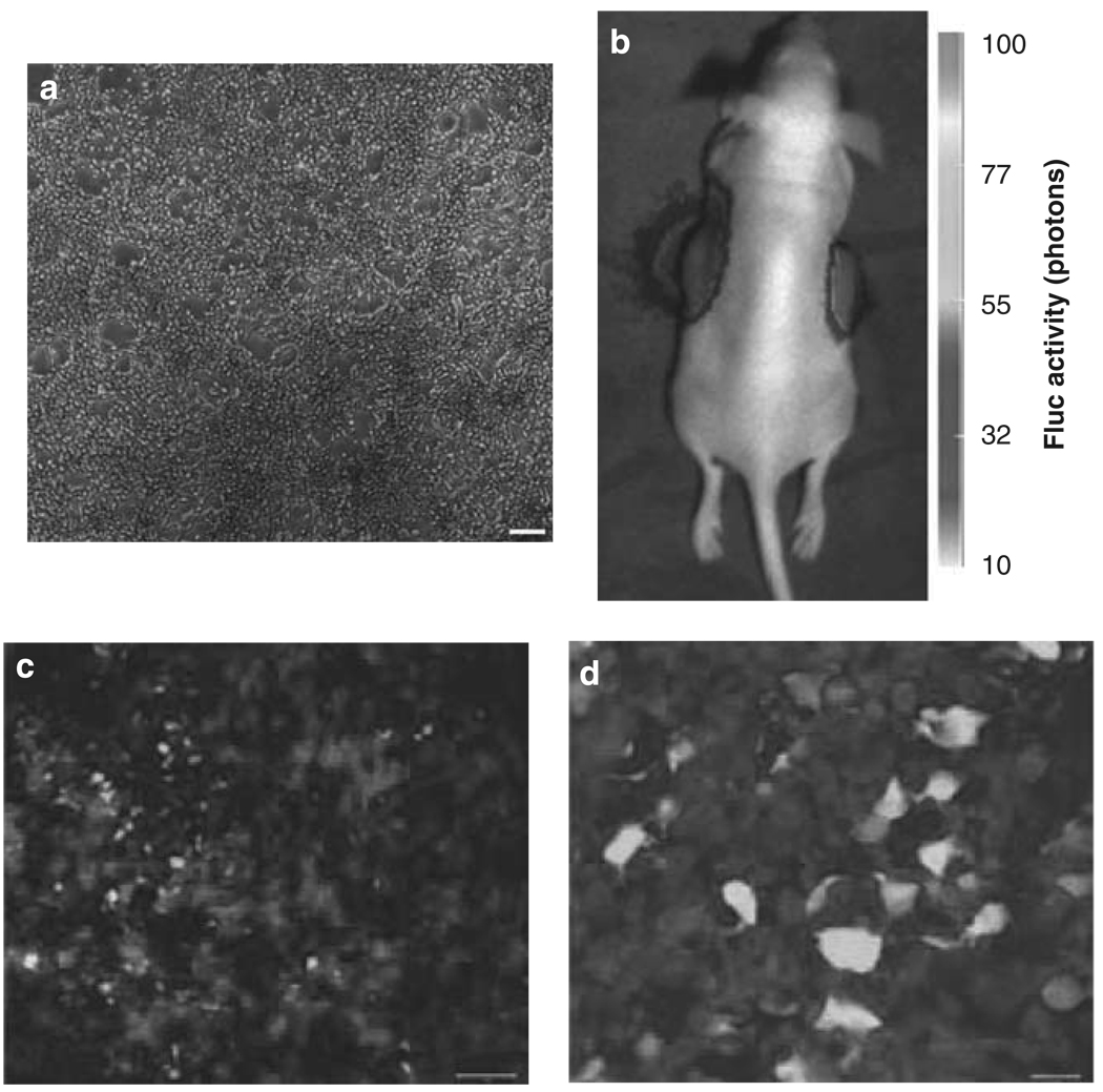
Figure 1.
Infectability of schwannomas in culture and in vivo with HSV amplicon vector. (a) HEI-193 cells were infected with HGC-Fluc HSV amplicon vector. Forty-eight hours later, cells were monitored for GFP fluorescence. (b) Five million HEI-193 cells were implanted subcutaneously into both flanks of nude mice. After 1 week, tumors were injected with 15 µl 107 tu HGC-Fluc vector. Twenty-four hours later, mice were injected with d-luciferin and imaged for Fluc activity using a CCD camera. (c–d) Tumors in (b) were removed 48 h post-injection and processed for GFP fluorescence (green) and S100 immunofluorescence staining (red). Scale bars: a, 100 µm; b, 20 µm. (See online version for color figure)

