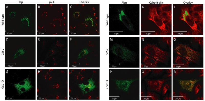
Figure 6. The G85V and G591D mutations lead to defective localization of ATP7B to the endoplasmic reticulum.
HEK293T cells were transfected with ATP7B-Flag (A–C and J–L), ATP7B-G85V-Flag (D–F and M–O) or ATP7B-G591D-Flag (G–I and P–R) and analyzed using antibodies against ATP7B (A, D, G, J, M, and P; green), p230 (B, E, and H; red) or Calreticulin (K, N, and Q; red), visualized by Alexa488 conjugated donkey anti-rat, Alexa568 conjugated donkey anti-mouse, Alexa568 conjugated donkey anti-rabbit IgG respectively. Overlap in staining is depicted in yellow in the overlay images (C, F, I, L, O, and R).

