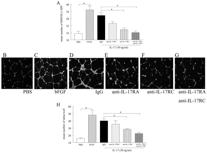
Figure 5. IL-17-mediated HMVEC chemotaxis and tube formation are regulated through both IL-17RA and IL-17RC.
A. HMVECs were incubated with mouse anti-human IL-17RA and IL-17 RC antibodies (10μg/ml) or control IgG (10μg/ml) for 1h. Thereafter HMVEC chemotaxis was performed in response to IL-17 (50 ng/ml) for 2h. PBS was used as a negative control, and VEGF (60 nM) as a positive control. HMVECs were incubated with antibodies to IL-17RA, IL-17RC, both IL-17RA and RC or IgG for 45 minutes at 37°C. Cells were then added to polymerized matrigel, IL-17 (50ng/ml), placed in the wells, and the plate was incubated for 16h at 37°C (in triplicate). Photomicrographs taken of representative wells treated with PBS (B), FGF (20 ng/ml) (C), IL-17 (50 ng/ml) plus IgG (D), IL-17 (50 ng/ml) plus anti-IL-17RA (10μg/ml) (E), IL-17 (50ng/ml) plus anti-IL-17RC (10μg/ml) (F) and IL-17 (50ng/ml) plus anti-IL-17RA and RC (10μg/ml) (G) in which IL-17-induced tube formation is significantly reduced by the neutralization of IL-17RC or both receptors (p<0.05). H. Data presented demonstrates mean number of branch points/tubes in each treatment group. Values are the mean ± SE, n=3. * represents p <0.05.

