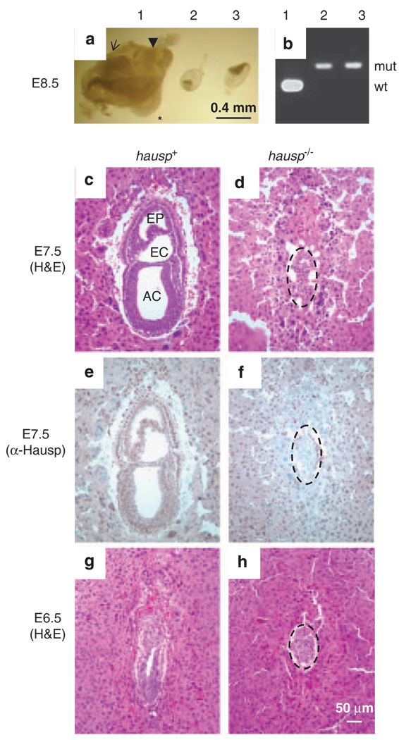
Figure 3.
Phenotypes of hausp knockout embryos. (a) Embryos at day E8.5 from the hausp heterozygote intercross are shown. Embryo no. 1 shows head fold (arrow), somites (*) and tail (arrow head), which are typically present in wild-type embryos at this stage; embryo nos 2 and 3 were abnormal, showing severely reduced cell mass and no advanced structures. (b) Genotyping results for the embryos showed in panel a, embryo no. 1 was wild-type and embryo nos 2 and 3 were hausp knockout embryos. (c) Deciduae from earlier developmental stages were analyzed by histology and immunostaining. Normal embryos at day E7.5 showed development of cavities, such as amniotic cavity (AC), exocoelom (EC) and ectoplacental cavity (EP). (d) Abnormal embryo at day E7.5 failed to develop any noticeable structures. Embryonic cell numbers were greatly reduced (dotted circle). (e) Genotyping of embryos from early embryonic development was performed by immunostaining using a polyclonal antibody anti-Hausp C-terminus. Embryonic cells from the embryo shown in panel c showed brown nuclear staining, suggesting it was either a wild-type or a hausp heterozygote embryo. (f) Embryonic cells (dotted circle) from panel d showed no Hausp staining, suggesting it was a hausp knockout embryo. The surrounding cells from the deciduae were stained by the Hausp antibody, as they were derived from a hasup heterozygote mother. (g) Wild-type embryos at E6.5 show formation of proamniotic cavity, and (h) hausp knockout embryo at E6.5 is smaller and delayed in development. Dotted circles in panels d, f and h, outline the hausp knockout embryos.

