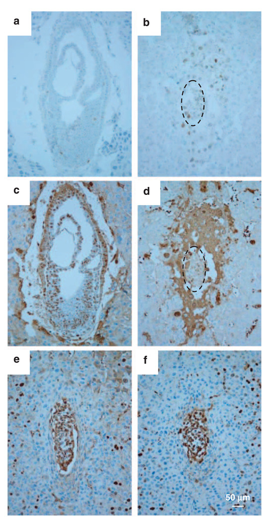
Figure 4.
Activation of p53 and assay of proliferative activity by BrdU incorporation. Activation of p53 was detected by immunostaining using anti-p53 polyclonal antibody. (a) The wild-type embryos at E7.5 from showed a few p53-positive cells, in contrast, (b) almost all embryonic cells were positive for p53 in hausp knockout embryo. Proliferative activity in embryos was determined by BrdU staining after BrdU incorporation. (c) More than 70% of cells in wild-type embryos at day E7.5 showed BrdU-positive staining, whereas (d) hausp knockout embryos had markedly reduced number of BrdU-positive cells, which was ~35%. However, BrdU-positive cells in wild-type embryo (e) and hausp knockout embryos (f) from day E6.5 were comparable. Dotted circles in panels b and d outline the hausp knockout embryos. BrdU, 5-bromo-2-deoxyuridine.

