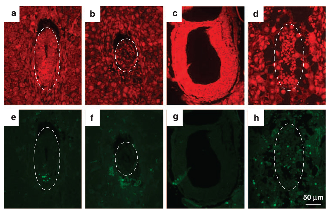
Figure 5.
Detection of apoptotic cells by TdT-mediated dUTP Nick-End Labeling (TUNEL) assay. Embryos from hausp heterozygote intercross were fixed and sectioned. The apoptotic cells were detected by TUNEL, followed by fluorescence microscopy (e–h). Nuclei were counter stained red by propidine iodide (a–d). Wild-type embryos are shown in panels a and e from day E6.5 and in panels c and g from day E7.5. Hausp knockout embryos are shown in panels b and f from day E6.5 and in panels d and h from day E7.5. There were minimum numbers of apoptotic cells detected in both wild-type embryos and hausp knockout embryos. Only wild-type embryo at day E6.5 (e) showed some apoptotic cells, which was likely normal during embryonic development. Dotted circles outline the embryos.

