
Fig. 1.
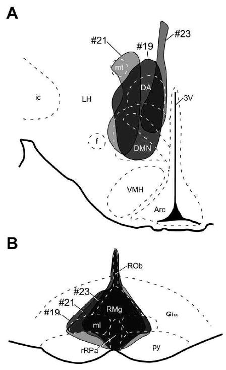
Sites of CTb injections. (A) Alexa488-conjugated CTb and Alexa594-conjugated CTb were injected into the DMH (A; bregma, −3.30 mm) and into the rRPa (B; bregma, −11.80 mm), respectively. Areas where injected CTb spread are shown on the brain maps adopted from an atlas of Paxinos and Watson (1998). Injections in the three animals (#19, #21 and #23) that were used for histochemical analyses are shown. 3V, Third ventricle; Arc, arcuate nucleus; DA, dorsal hypothalamic area; DMN, dorsomedial hypothalamic nucleus; f, fornix; Giα, alpha part of the gigantocellular reticular nucleus; ic, internal capsule; LH, lateral hypothalamic area; ml, medial lemniscus; mt, mammillothalamic tract; py, pyramidal tract; ROb, raphe obscurus nucleus; RMg, raphe magnus nucleus; VMH, ventromedial hypothalamic nucleus.