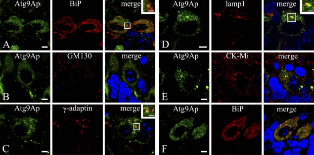
Figure 4.
Localization of Atg9Ap in Purkinje cells of the cerebellum in mice at 8 (A–E) and 3 (F) weeks of age. (A–F) Mouse Purkinje cells were coimmunostained with the anti-Atg9A antibody (green) and with an antibody for various organelle markers (red): BiP for endoplasmic reticulum (A,F); GM130 for Golgi apparatus (B); γ-adaptin for TGN (C); lamp1 for lysosome/late endosome (D); and mitochondrial creatine kinase (CK-Mi) for mitochondria (E). Nuclei were counterstained with 4′,6-diamidino-2-phenylindole. Bars: A–F = 5 μm; insets = 2 μm.

