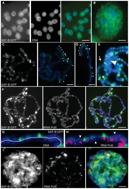
Figure 5. Distribution of SAF-B-GFP in polytene larval salivary gland nuclei.
(A) Immunodetection of SAF-B-GFP, DAPI staining of DNA, and the merge. The same nucleoplasmic and focal distribution seen in diploid cells are apparent. Additionally, more intense foci and threadlike continua are seen to connect foci. (B) Increased magnification to show continua. Brighter foci are at confluences of continua. (C) Squashed nuclei showing distribution of SAF-B-GFP, and merge with DAPI-stained DNA. SAF-B associates with specific bands on polytene chromosomes. White dots highlight SAF-B bands at the tip of the X chromosome. (D) A different polytene X chromosome, labelled as in (C), showing consistency of banding pattern. (E) The chromocenter (arrowhead) of salivary gland chromosomes does not show enhanced or reduced localization of SAF-B-GFP, although some foci are seen, and the nucleolus (arrows) shows only very low level of SAF-B-GFP. (F) Immunodetection of SAF-B-GFP, RNA Polymerase II (Ser2-PO4), and merge with DAPI-stained DNA. Extensive areas of overlap of both epitopes are evident, as are bands with detection of only SAF-B-GFP or RNA Polymerase II. (G) Immunodetection of SAF-B-GFP counterstained with DAPI to reveal salivary gland chromosome bands. SAF-B localized primarily to interband regions. (H) Immunodetection of SAF-B-GFP and DAPI staining of DNA as in (G), with immunodetection of RNA Polymerase II (Ser2-PO4), showing most bands of SAF-B overlap with RNA Polymerase II, but some bands of only one detectable epitope are apparent (arrowheads). (I) Immunodetection of SAF-B-GFP and RNA Polymerase II (Ser2-PO4) in whole-mount salivary gland nuclei, and merge with DAPI-stained DNA. Scale bar 50 µm (A), 10 µm (B–E, I), or 20 µm (F).

