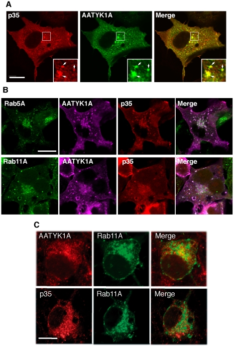
Figure 2. Colocalization of p35 with AATYK1A in early and recycling endosomes.
(A) Colocalization of AATYK1A and p35 in COS-7 cells. COS-7 cells were transfected with AATYK1A-Myc together with p35 and Cdk5. AATYK1A and p35 were detected by immunostaining with the anti-Myc antibody and anti-p35 antibody, followed by incubation with Alexa 488-conjugated anti-mouse IgG and Alexa 548-conjugated anti-rabbit antibody, respectively. A merged image is shown in the right panel. Insets represent higher magnifications and arrows indicate the colocalization. Scale bar, 20 µm. (B) Localization of AATYK1A and p35 in early and recycling endosomes. COS-7 cells were transfected with AATYK1A-Myc, p35, Cdk5, and either EGFP-Rab5A (as an early-endosome marker) or EGFP–Rab11A (as a recycling-endosome marker). After 24 h of transfection, cells were fixed and stained with anti-Myc and anti-p35 antibodies, as described above, and were observed using a confocal microscope. Scale bar, 10 µm. (C) Localization of AATYK1 and p35 in endosomes in cultured cortical neurons. Rat brain cortical neurons at DIV5 were transfected with EGFP-Rab11A (middle panels). The cells were immunostained with anti-AATYK1 and p35 (C19) 24 h after transfection, followed by Alexa 546-conjugated anti-rabbit secondary antibody (left panels). Merge is shown in right panels. Bar, 10 µm.

