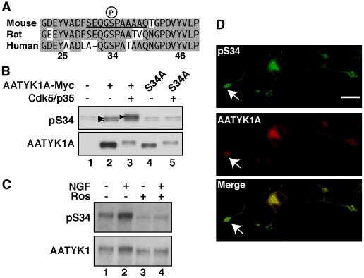
Figure 4. Generation of the anti-pSer34-specific antibody and Ser34 phosphorylation of AATYK1 in PC12D cells.
(A) Amino-acid sequences of mouse, rat, and human AATYK1A around Ser34. A synthetic peptide corresponding to the mouse AATYK1A amino-acid residues 29–39 (the Ser34 phosphorylation site is underlined) was used for rabbit immunization. (B) Specificity of the anti-pS34 antibody. COS-7 cells were transfected with AATYK1A or its S34A mutant in the presence (+) or absence (–) of Cdk5/p35. Cell extracts were immunoblotted with the anti-pS34 antibody or anti-Myc antibody for AATYK1A. (C) Phosphorylation of AATYK1 at Ser34 in PC12D cells. PC12D cells were treated with 50 ng/ml NGF for 24 h in the presence or absence of 20 µM roscovitine (Ros). AATYK1 was immunoprecipitated in PC12D cells using the anti-AATYK1 antibody and was subjected to immunoblotting with the anti-pS34 or anti-AATYK1 antibodies. (D) Immunofluorescent staining of PC12D cells using the anti-pS34 antibody. PC12D cells expressing AATYK1A-Myc were treated with NGF for 24 h and double labeled with the anti-pS34 (top panel) and anti-Myc (AATYK1A, middle panel) antibodies. A merged image is shown in the lower panel. The growth cone is indicated by an arrow. Scale bar, 20 µm.

