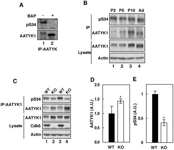
Figure 5. Cdk5-mediated in vivo Ser34 phosphorylation.
(A) Phosphorylation of AATYK1 in mouse brain. AATYK1 was immunoprecipitated from mouse brain extracts and incubated in the presence (+) or absence (–) of bacterial alkaline phosphatase (BAP). The anti-pS34 reaction is shown in the upper panel and the anti-AATYK1 reaction is shown in the lower panel. (B) Phosphorylation of AATYK1 in mouse brain during early postnatal development. AATYK1 was immunoprecipitated from mouse brain extracts at P2, P5, P10, and six weeks of age (Ad) using the anti-AATYK1 antibody. The immunoprecipitates were immunoblotted using the anti-pS34 (top panel) and anti-AATYK1 (second panel) antibodies. Immunoblots of brain extracts using anti-AATYK1 and anti-actin antibodies are also shown in the lower panels. (C) Phosphorylation of AATYK1 at Ser34 in Cdk5–/– mouse brain. AATYK1 was immunoprecipitated from brain extracts of Cdk5–/– mice at embryonic day 18.5 (E18.5) and immunoblotted using the anti-pS34 antibody (top panel). Immunoblots of the brain extracts using anti-AATYK1 (third panel), anti-Cdk5 (fourth panel), and anti-actin (bottom panel) antibodies. Quantification of AATYK1 and pS34 is shown in (D) and (E), respectively. Bars indicate the means ± S.E. of three independent experiments (n = 3; * P<0.05; t test).

