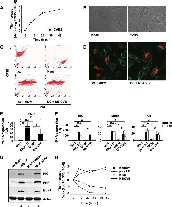
FIG. 2.
Min6 cells are a good model for primary pancreatic cells, are phagocytosed by DCs, and induce innate antiviral immune responses. A and B: Min6 cells were infected with CVB3 at an MOI of 10, and replication was analyzed at indicated time points (A) and images were taken at 48 h postinfection (p.i.) (B). C: Min6 cells were PKH-labeled and infected with CVB3 at an MOI of 10. After 24-h incubation, cells were harvested and added to CFSE-labeled DCs at a 1:1 ratio. After 24-h co-culture, cells were harvested and uptake was determined using flow cytometry. D: Min6 cells were prepared as in C and co-cultured with unlabeled DCs for 24 h, after which DCs were harvested, stained as in Fig. 1E, and analyzed using confocal microscopy. E and F: Min6 cells were prepared as in C, and after 48-h incubation, cells were harvested and added to DCs at a 1:1 ratio or DCs were stimulated with 20 μg/ml poly (I:C) or left untreated. At 5 h after addition (E), or 5 h and 8 h after addition (F), mRNA induction of ISGs was determined as described. G: Protein expression of RIG-I, Mda5, and PKR was analyzed by Western blot 24 h after stimulation of DCs as described for panel E. H: DCs were stimulated as in E, and after 24-h co-culture cells were harvested and infected with EV9 at an MOI of 1. At indicated times postinfection, EV9 replication was analyzed. Poly (I:C) was used as a positive control (34). In some experiments, freeze-thawed cell populations were used but these yielded similar results compared with using viable cells (supplemental Fig. 1). Data shown are representative (A–D, G, and H) or averages (E and F) of at least three independent experiments. Med, medium, i.e., unstimulated cells; M6/CVB, CVB-infected Min6 cells; M6/M, mock-infected Min6 cells; n.s., not significant. *P < 0.05. (A high-quality color representation of this figure is available in the online issue.)

