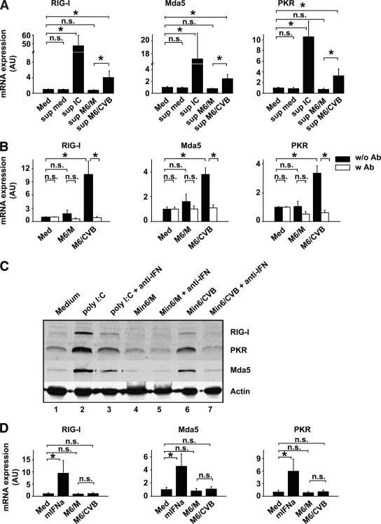
FIG. 3.
Type I IFNs produced by DCs themselves are required for ISG induction. A: DCs were stimulated with cleared supernatants from stimulated DC and DC/Min6 co-cultures (harvested 24 h after co-culture started and used at a 1:2 dilution) and mRNA induction of RIG-I, Mda5, and PKR were determined using qPCR 8 h after stimulation. B: Min6 cells were infected with CVB3 at an MOI of 10 and incubation cells were harvested and added to DCs at a 1:1 ratio after 48 h. Stimulations were performed in the absence or presence of neutralizing antibodies (Iivari, Kaaleppi, and bovine anti–IFN-α/β; see research design and methods). After 8 h, mRNA expression levels of RIG-I, Mda5, and PKR were determined using qPCR. C: DCs were treated as in B and protein expression of RIG-I, Mda5, and PKR was analyzed by Western blotting after 24 h. D: DCs were stimulated with 100 units/ml mIFNα or cleared supernatants from Min6 cells (harvested 48 h postinfection and used at a 1:2 dilution), and ISG mRNA induction was determined after 8 h. Data shown are representative of two (C) or average of three (A, B, and D) independent experiments. IC, poly (I:C); Med, medium, i.e., unstimulated cells; M6/CVB, CVB-infected Min6 cells; M6/M, mock-infected Min6 cells; mIFNa, murine recombinant IFN-α; n.s., not significant; Sup, supernatant; w/o Ab or w Ab, without or with neutralizing anti–IFN-α/β antibodies, respectively. *P < 0.05.

