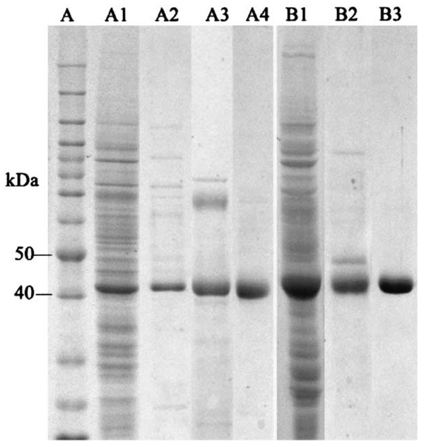
Fig. 1.
Recombinant Ae-HKT/AGT and Dm-Spat expression and purification. Soluble proteins were obtained as described in Section 2 and subjected to SDS–PAGE analysis. Lanes (left side) illustrate protein profiles of crude soluble proteins from cells infected with the Ae-HKT/AGT recombinant virus (A1), the remaining proteins after DEAE Sepharose (A2), phenyl Sepharose (A3) and hydroxyapatite (A4) chromatographic separations, respectively. Lanes (right side) represent soluble protein from cells infected with the Dm-Spat recombinant virus (B1), the remaining protein after phenyl Sepharose (B2) and hydroxyapatite (B3) chromatographic separations, respectively. Lane A shows the protein molecular mass standards.

