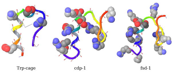
Figure 6.
Graphical representations of the NMR structures of the three miniproteins investigated in this work: trp-cage (pdb id: 1RIJ), cdp-1 (pdb id: 1PSV), and fsd-1 (pdb id: 1FSD). In each case the first deposited NMR model is shown. Backbone ribbon is colored from N-terminal (red) to the C-terminal (blue). Charged sidechains are shown in space-filling representation.

