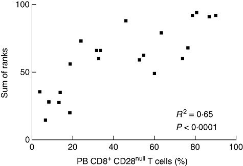
Fig. 6.

Scatter diagram illustrating the correlation between the percentage of peripheral blood (PB) CD8+CD28null T cells and the sum of ranks of the individual significant correlations with the percentage of bronchoalveolar lavage fluid (BALF) CD4+CD28+, CD4+ very late antigen (VLA-1)+, CD8+CD25+, CD8+VLA-1+ and CD8+ human leucocyte antigen D-related (HLA-DR)+ lymphocytes from sarcoidosis patients.
