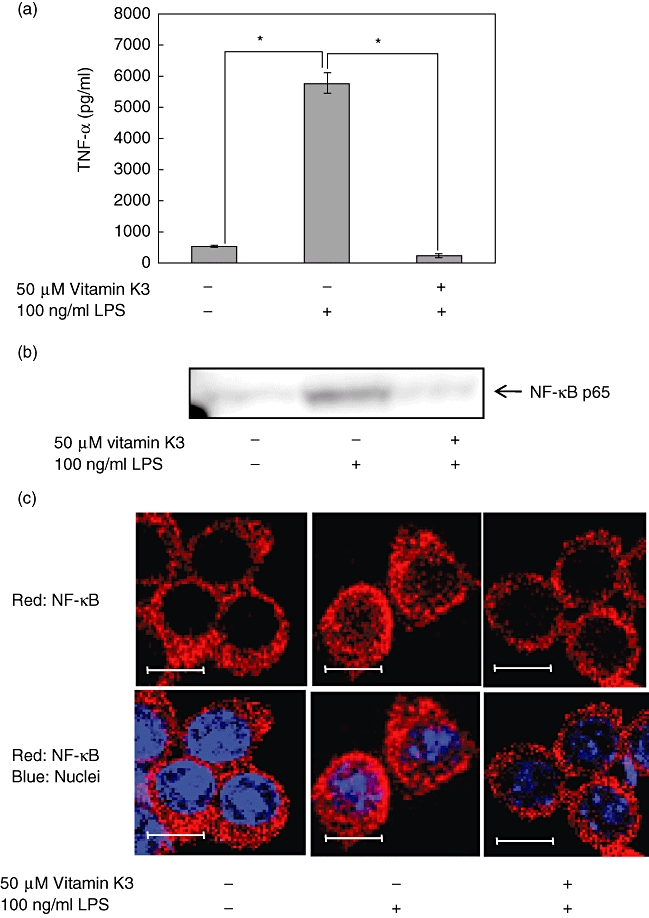
Fig. 3.

Inhibitory effects of vitamin K3 on the lipopolysaccharide (LPS)-induced tumour necrosis factor (TNF)-α production and nuclear factor (NF)-κB nuclear translocation in RAW264·7 cells. (a) RAW264·7 cells were pretreated with 50 µM vitamin K3 or DMSO as a vehicle control for 30 min and then treated with 100 ng/ml LPS for 24 h, and the TNF-α concentration in the cell medium was measured by enzyme-linked immunosorbent assay. Each data value is expressed as mean ± standard error of duplicates of three experiments. The presence of significant differences is indicated by asterisks (P < 0·05). (b) RAW264·7 cells were incubated with 50 µM vitamin K3 or dimethylsulphoxide (DMSO) as a vehicle control for 30 min, followed by treatment with 100 ng/ml LPS for 30 min. The nuclear proteins were prepared from the cells and were subjected to Western blot analysis for evaluation of the nuclear translocation of NF-κB p65. Typical pictures are shown from at least triplicate determinations. (c) RAW264·7 cells were treated as described in Fig. 3b, and the nuclear translocation of NF-κB p65 was assessed by immunofluorescence microscopy (red; NF-κB; blue: nuclei). Typical pictures are shown from at least triplicate determinations. Scale bar, 10 µm.
