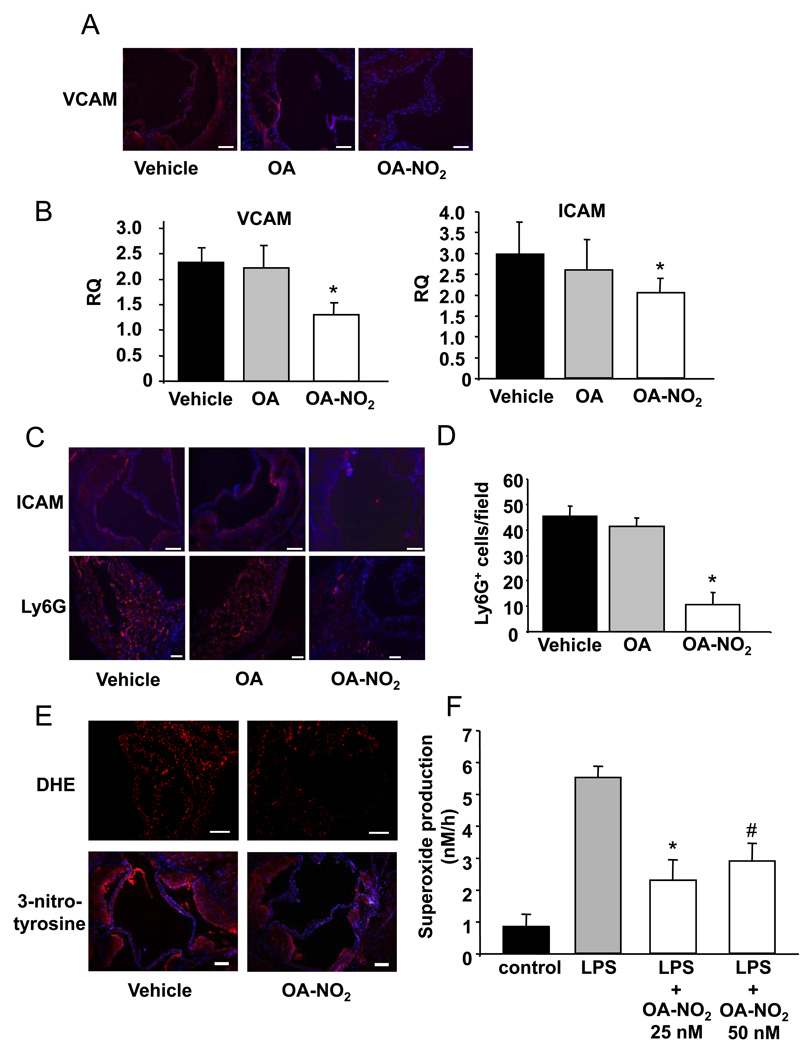
Figure 4.
Vascular and intracellular adhesion molecules were down-regulated by OA-NO2. A, Representative immunostained aortic sections using specific antibodies against VCAM (Magnification 20X, scale bar indicates 100 µm; blue=nuclei, red=VCAM). B, VCAM and ICAM mRNA expression in the aortic wall was lower in OA-NO2-treated animals (p<0.05, *p<0.05 vs. vehicle and vs. OA). C, Representative immunostained aortic sections using ICAM and Ly6G antibodies showed less ICAM expression and neutrophil infiltration in OA-NO2-treated animals (Magnification 20X, scale bar indicates 100 µm, blue = nuclei, red = ICAM/neutrophils). D, Densitometric quantification of Ly6G positive cells per field of view (p<0.001, *p<0.001 vs. vehicle and vs. OA). E, Representative immunostaining for DHE (Magnification 20X, scale bar indicates 100 µm) and 3-nitrotyrosine (Magnification 10X, scale bar indicates 100 µm, blue=nuclei, red=3-nitrotyrosine). F, Ex vivo LPS-induced superoxide production by BMDMs isolated from apoE−/− mice with and without OA-NO2 treatment (p=0.004, *p=0.003 vs. LPS, #p=0.07 vs. LPS).

