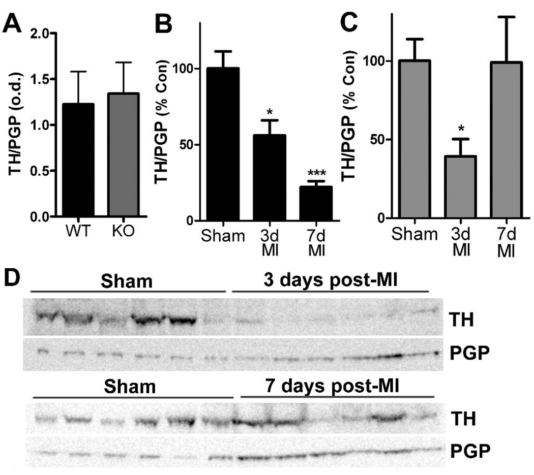
Figure 2.
Tyrosine hydroxylase content in the left ventricle. TH was quantified by western blot and normalized to the pan-neuronal marker PGP. A) Neuronal TH content is identical in the left ventricle of WT and gp130 KO mice. B) Neuronal TH content in WT mice declined 3 days after MI, and decreased further 7 days after ischemia-reperfusion surgery. Data shown are the Mean±SEM; n=6; * p<0.05, *** p<0.001. C) Neuronal TH content in gp130 KO mice decreased 3 days after MI but returned to control levels 7 days after ischemia-reperfusion. Data shown are the Mean±SEM; n=6; * p<0.05. D) Representative western blots showing TH and PGP in gp130 KO left ventricle from 6 sham and 6 post-MI mice.

