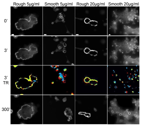
Fig. 5.
Frames from a time-lapse movie of the sealing zones of osteoclasts on rough and smooth calcite surfaces conditioned with 5 μg/ml and 20 μg/ml vitronectin, viewed at 0, 3 and 300 minutes. The initial time point is arbitrary. Temporal ratio (TR) images are color-coded, as described for Fig. 2. Note that the dynamics, size and number of sealing zone rings depend on the topography of the calcite surface, regardless of the amount of vitronectin adsorbed. Scale bars: 10 μm.

