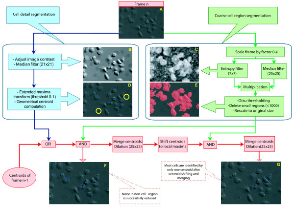
Figure 2.
Cell centroid segmentation. Schematic workflow and examples of intermediary steps of cell centroid extraction from microscopic images. Each new frame (A) will be processed in two distinct steps, namely cell detail segmentation (left, blue box) and cell region segmentation (right, green box). The detected centroids from the detail segmentation are first combined with the extracted centroids of one past frame to propagate cell centroids steadily through an image sequence. Afterwards the combination of the cell region image and the cell centroid image leads to deletion of cell positions in non-cell regions (panel F). Subsequent centroid merging and shifting finally concentrate groups of possible centroids within one cell to form a single cell centroid (panel G).

