
Figure 6.
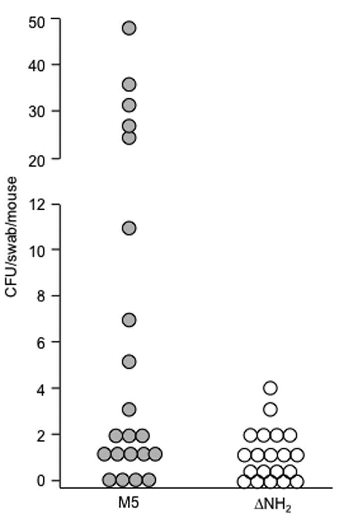
Colonization of the upper respiratory tract of mice infected intranasally with M5 and its ΔNH2 mutant. Mice were infected intranasally with wt M5 (closed circles) and ΔNH2 mutant (open circles) grown in THB overnight, washed and resuspended to an O.D. of 1.00. Six hours after inoculation the oral cavity of each mouse was swabbed and the swabs were plated onto blood agar. The number of beta-hemolytic colonies was recorded after 24 hours incubation. There was a significant difference in colonization between wt M5 (mean 9.97+/− 3.1 CFU, median 1 CFU) and ΔNH2 mutant (mean 1.0+/−0.26 CFU, median 0 CFU) infected mice (p<0.05, Wilcoxon test).