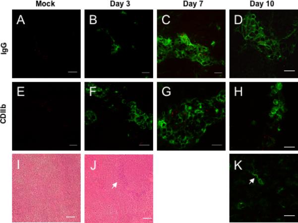
Figure 5.

CPMV penetrates the BBB during CNS infection. A-D) Fluorescence immunohistochemistry with A488 conjugated anti-mouse IgG in the brains of mock infected mice and on days 3, 7, and 10 after MHV infection. E-I) FITC conjugated anti-CD11b staining in mouse brains after mock infection and days 3, 7, and 10 after intracerebral inoculation with MHV. Red = CPMV. CDllb+ microglial cells exhibit a characteristic stellate morphology and arrow indicates colocalization of a microglial cell with CPMV (I). Magnification is 600x and scale bar is 15 microns. H &E staining of brain parenchyma from mock-infected (I) and MHV-infected (J) mice. Arrow indicates inflammatory cells within the tissue. Images are representative of three mice examined at each timepoint.
