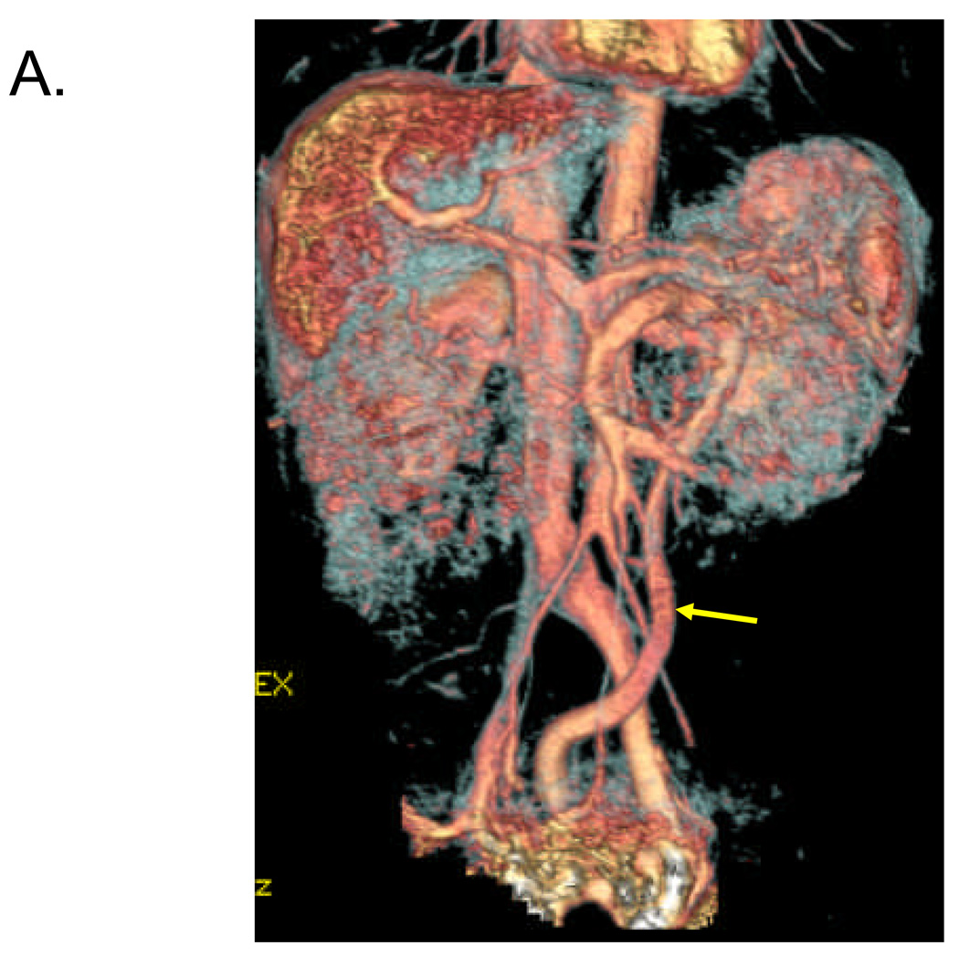
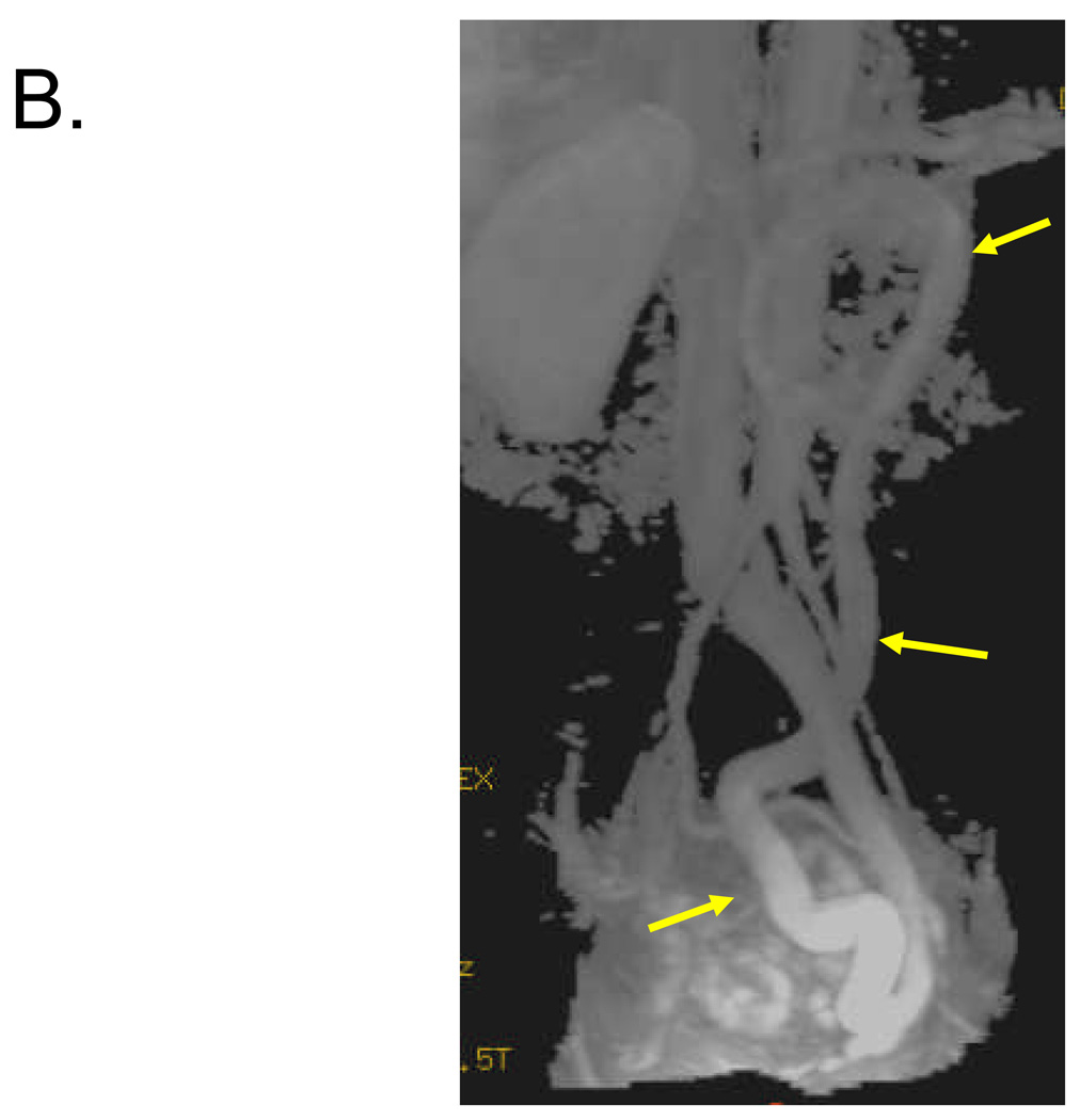
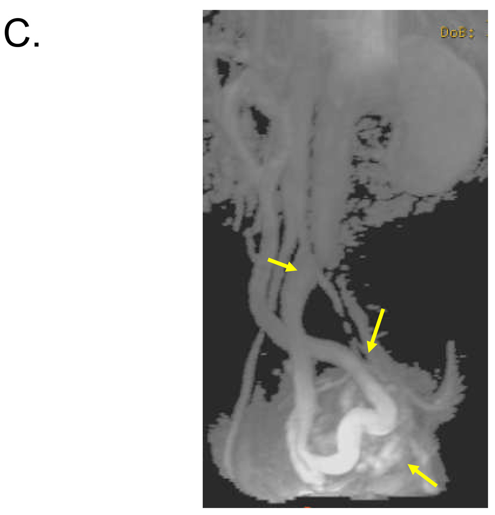
Figure 1. Identification of 1–2 cm shunt between SMV and left internal iliac vein in 57 year old female.
Volume rendered (A) and maximum intensity projection (B, C) reconstructed images from venous phase of contrast-enhanced 3D MRA demonstrate a long serpiginous shunt (arrows in A, B and C) which arises from the SMV near the portal confluence, descends into the pelvis, and empties into the left internal iliac vein.


