
Figure 2. Effect of IL-1β on osteoblasts and stem cells in vitro.
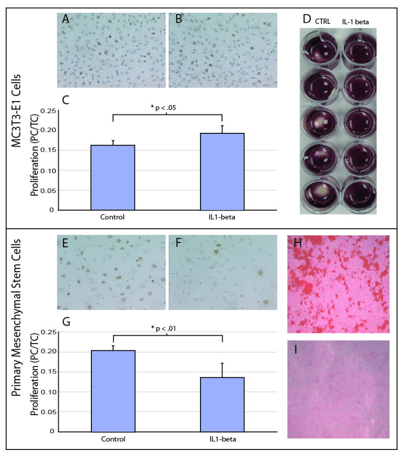
(A,C) Proliferation of control pre-osteoblasts (n=5), and (B,C) pre-osteoblasts cultured with IL-1β (n=5, PC/TC=proliferating cells to total cells). Proliferating cells are stained with BrdU staining. (D) Differentiation of preosteoblasts in the presence (n=5) and absence (n=5) of IL-1β. Cells are stained with Alizarin red. (E,G) Proliferation of control MSCs (n=4) and (F,G) MSCs cultured with IL-1 β (n=5). Proliferating cells are stained with BrdU staining. (H) Differentiation of control MSCs (n=5), and (I) MSCs in the presence of IL-1 β (n=5). Cells are stained with Alizarin red. Data are mean +/-standard deviation.