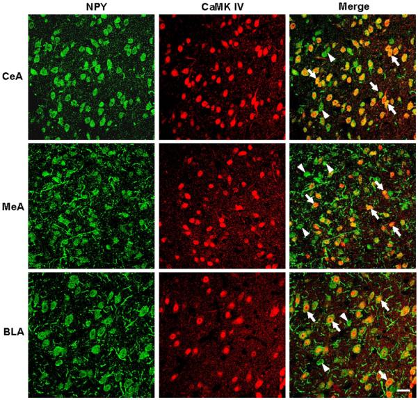
Figure 6.
Representative photomicrographs showing the immunofluorescence staining for NPY (green) and CaMK IV (red) in the cells of CeA, MeA and BLA. The colocalization of the NPY and CaMK IV in cells is indicated by the yellow color development (merge). These photomicrographs show that most of the NPY-positive cells co-express CaMK IV (arrows) with a few exceptions (arrowheads). Scale bar = 20 μm.

