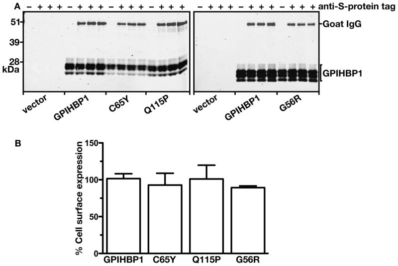
Figure 4. Assessing the amounts of GPIHBP1 at the surface of cells relative to the total amounts of GPIHBP1 in the cell with a goat antibody against the S-protein tag and a mouse monoclonal antibody against human GPIHBP1.
(A) Western blot analysis of GPIHBP1 at the cell surface. CHO pgsA-745 cells were electroporated with an empty vector or an expression vector encoding wild-type human GPIHBP1, GPIHBP1-C65Y, GPIHBP1-Q115P, or GPIHBP1-G56R. All constructs contained an amino-terminal S-protein tag. The next day, the cells were incubated for 2 h at 4° C with a goat antiserum against the S-protein tag. After washing the cells six times in ice-cold PBS, cell extracts were prepared for western blotting with a IR680-conjugated donkey antibody against goat IgG and a mouse monoclonal antibody against human GPIHBP1. The mouse monoclonal antibody was detected with an IR800-conjugated donkey antibody against mouse IgG.
(B) Quantification of the western blot. The signal corresponding to the goat anti-S-protein tag IgG was normalized to the signal for the monoclonal antibody against human GPIHBP1 and expressed relative to the ratio for wild-type GPIHBP1 (set at 100%).

