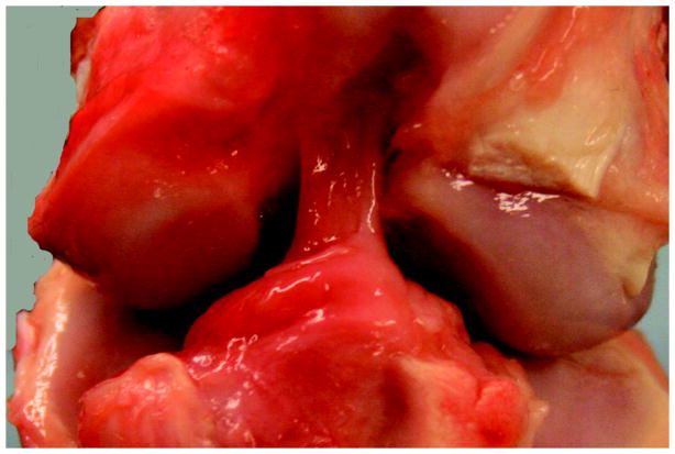
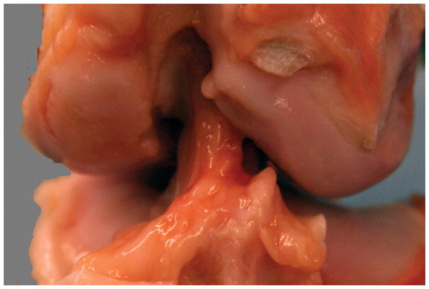
Figure 3.
Figure 3A: Gross appearance of a knee treated with SUTURE repair alone. Note the repair tissue arising from just posterior to the fat pad, from the anterior slope of the tibial spines, and coursing from the tibia to the lateral wall of the intercondylar notch. There is no significant change noted in the femoral condylar cartilage either medially or laterally. The repair tissue itself is composed of multiple individual synovialized bands.
Figure 3B. Gross appearance of a knee treated with SCAFFOLD supplementation of the suture repair. Note the similar appearance with the SUTURE knee in 3A. Again, there is repair tissue coursing from tibia to femur in one continuous mass. There is no change noted in the femoral condylar cartilage. The repair tissue itself is composed of multiple individual synovialized bands.


