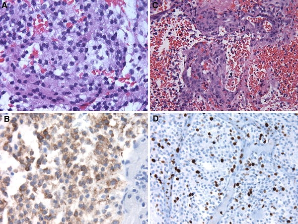
Fig. 2.

a Microscopic evaluation showed an atypical neurocytic tumor composed of cells with round to oval nuclei. (H&E 400×). b Diffuse, strong immunolabeling for synaptophysin was demonstrated. (400×). c Atypical features were noted including prominent glomeruloid vascular proliferation (H&E 400×). d The Ki-67 labeling index was approximately 10% overall (400×)
