Fig. 4.

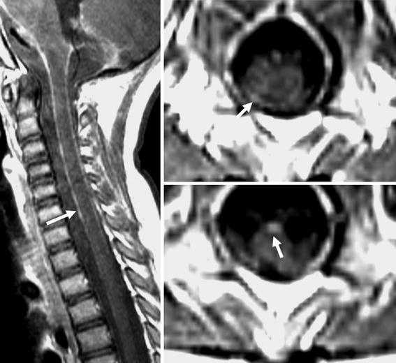
Post contrast sagittal T1 and axial T1-weighted images show enhancement of the surface of the cord (white arrow), suggesting recurrent disease

Post contrast sagittal T1 and axial T1-weighted images show enhancement of the surface of the cord (white arrow), suggesting recurrent disease