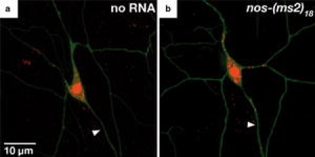
Fig. 2.

Imaging of nos RNA in Drosophila peripheral larval neurons. Class IV dendritic arborization (da) neurons in semi-intact third instar larvae expressing a MCP-RFP (red) alone (control) or b MCP-RFP (red) and nos-(MS2)18 mRNA (Brechbiel and Gavis 2008); Unbound MCP-RFP is sequestered in the nucleus due to a nuclear localization signal (NLS). Arrowheads indicate the axon. Neurons are filled with GFP (green). Pictures are reproduced with permission from © Brechbiel and Gavis (2008). Originally published in Current Biology. doi:10.1016/j.cub.2008.04.033
