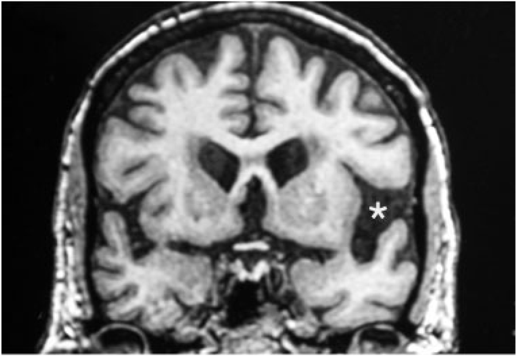
Fig. 1.
Coronal magnetic resonance imaging (MRI) scan of Case 8 done 1 year after onset. The left perisylvian cistern is wider (asterisk), indicating more atrophy on that side. Postmortem examination also showed asymmetry, with greater microvacuolation in the left neocortex. However, there were no asymmetries of neurofibrillary tangle (NFT) distribution. Visual inspection of the MRI also showed that perisylvian atrophy was more severe than medial temporal atrophy. However, the NFT density did not show a perisylvian over entorhinal predominance. This patient illustrates the inconsistency of correlation between atrophy patterns and NFT distribution in primary progressive aphasia/Alzheimer’s disease (PPA/AD).

