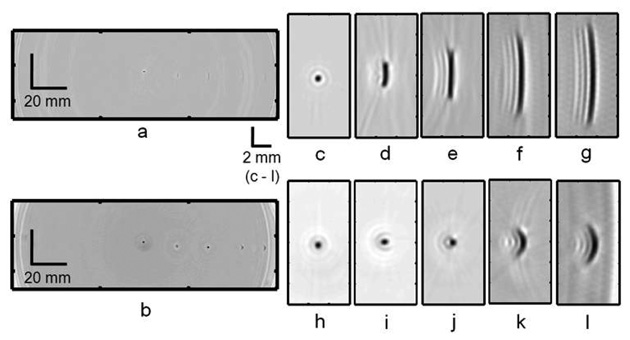
FIG. 3.
PAT images of five 0.5 mm diameter pencil leads placed inside the scanning region at different distances from the scanning center. (a) Reconstructed PAT image using the flat detector. (b) Reconstructed PAT image using the negative lens detector. (c) to (g) Close-up images of all five objects in (a) at distances of ~2 mm, ~19 mm, ~36 mm, ~55 mm, and ~67 mm from the scanning center, respectively. (h) to (l) Corresponding close-up images obtained with the negative lens detector.

