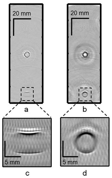
FIG. 6.
Reconstructed cross-sectional PAT images of two LDPE tubes (~1 cc volume, inner diameter ~6 mm) filled with diluted India ink, one placed near the scanning center and the other at a distance of ~50 mm from the scanning center. (a) Image using the flat detector. (b) Image using the negative lens detector. (c) Close-up image of the tube at ~50 mm from the scanning center using the flat detector. (d) Close-up image of the tube at ~50 mm from the scanning center using the negative lens detector.

