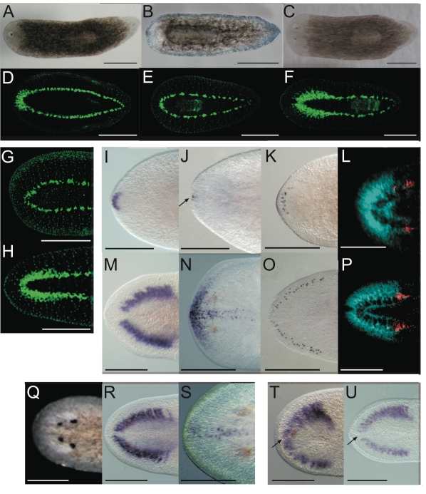
Figure 2. Smed-prep(RNAi) leads to the loss of anterior fate during regeneration.
Smed-prep(RNAi) using a standard injecting and cutting protocol (Figure S2A) leads to animals with either one (A) or no eyes (B). Control gfp(RNAi) animals were all normal (C). Staining with the 3C11 monoclonal antibody to synapsin in Smed-prep(RNAi) with one eye (D), animals with no eyes (E), and gfp(RNAi) (F). Smed-prep(RNAi) animals (Figure S2B) (G) and gfp(RNAi) injected during regeneration. Staining with a probe to a glutamate receptor specific to CG/brain, branches, Smed-GluR, confirms reduction of CG structure to the most anterior tip (I). Smed-sFRP-1, a marker of anterior fate, is mostly absent or else confined to the very anterior tip (J). Staining with cintillo (K) shows that the number of these anterior cells is also reduced and restricted to the anterior tips of animals. Staining with the posterior brain marker Smed-WntA (red) shows that in animals where CG/brain is present A/P polarity of the brain (DAPI stained in blue) is maintained (L,P). gfp(RNAi) were normal for all these stains (M–P). Prolonged Smed-prep(RNAi) during homeostasis (Figure S2C) leads to the formation of two new eyes anterior to the original pair (Q) but not to any visible reduction or incorrect patterning of the CG/brain, as shown by Smed-GluR expression (R). The most anterior margin expression of Smed-sFRP-1 is lost in Smed-prep(RNAi) homeostasis worms (S). Smed-prep(RNAi) worms amputated laterally (Figure S2A) are able to regenerate CG, as shown by Smed-GluR expression (T), but the regeneration is not patterned correctly as branches are fused (see arrow in T) compared to gfp(RNAi) animals (U). All panels depict 12 day regenerating trunks except: (G,H) 12 day regenerating tails, (Q,R,S) 28 days homeostasis after first injection, (T,U) 15 days regeneration after lateral regeneration. All scale bars 1 mm.

