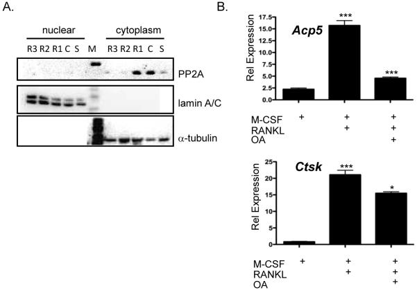
Figure 4. Expression of PP2A in osteoclasts.
(A) Representative immunoblot of lysates from bone marrow-derived osteoclasts were M-CSF and RANKL was removed for 4-6 h, stimulated with M-CSF or M-CSF and RANKL for up to 72 h and immunoblotted against the catalytic subunit of PP2A, α-tubulin and lamin A/C. (B) Real-time RT-PCR analysis of osteoclasts treated with or without 20 nM oakdaic acid 6 h before harvest for expression of Acp5 ***p<0.0002 vs. M-CSF treated, ***p<0.0005 vs. RANKL treated or cathepsin K, ***p<0.0001 vs. M-CSF treated, *p<0.01 vs. RANKL treated.

